
The Z-band (Z-line, Z-disc) is an intriguing structure that tethers actin filaments from adjacent sarcomeres into a tetragonal lattice. During contraction, the tension generated between the myosin and actin filaments is transmitted through the Z-band components to the adjoing sarcomere and so to the ends of the fibre.
To understand the 3D structure of the Z-band, we need to obtain different views by tilting the section about the filament axis (vertical here). The different views are combined by computer and a 3D reconstruction done by Fourier methods or by tomography. The following analysis is from my paper on 3D structure of the Z-band in fish fin muscle: Luther (2000).

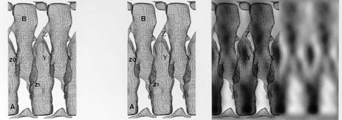
These are different lattice views of the Z-band in fish fin muscle.

Average lattice views from previous figure. We refer to these views as (a) [10], (b) [01], (c) [11] & (d) [1-1]. The Fourier transforms were combined and then 3D reconstruction was calculated.
3D reconstruction of Z-band in fish fin muscle; left, stereo view; right, superimposed on average lattice view.
Stereo view of 3D model of fish fin muscle Z-band, TS view.

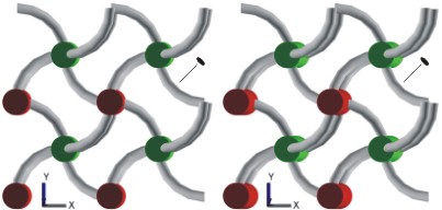
Stereo view of 3D model of fish fin muscle Z-band, [10] view. This Z-band comprises 3 layers of α-actinin (labelled Z0, Z1 and Z1').
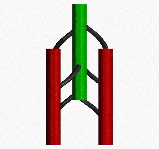
Movie showing the path of the 3 layers of α-actinin links (black) between actin filaments from adjacent sarcomeres