Transverse section of the bare regions in fish (left) and frog (right) striated muscles. Myosin filaments have triangular profiles. In fish, the triangles point the same way, but in frog they have two orientations that give rise to a superlattice arrangment (described below).

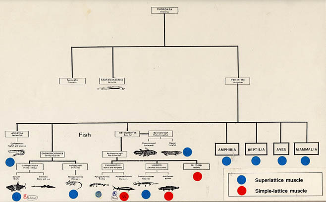
Distribution of superlattice and simple-lattice muscles in the vertebrates.